

Integrated Imaging Diagnostic & Education Centre
A new initiative to support your patients with geographic atrophy


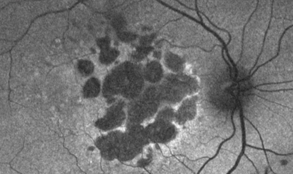
CERA, in collaboration with Pliny, has introduced a complimentary diagnostic imaging interpretation service designed to support eyecare professionals in distinguishing between geographic atrophy related to AMD and inherited retinal diseases. It is designed to support and educate clinicians regarding lesion characteristics pertaining to their diagnosis and guidance about when to refer or recommend treatment. Additionally, if any PBS lesion criteria are introduced for upcoming treatments, IIDEC will provide feedback on these criteria.1
A first for Australia. A step forward for eye care.
This is the first foray into remote analysis of raw OCT data in Australia and sets a precedent that will potentially lead to new ways of collaboration and management of a wide range of conditions.
.png)
“For the management of geographic atrophy, the correct diagnosis and determining which patients will benefit most from treatment, is key.”
Prof Robyn Guymer AM
Centre for Eye Research Australia
How it Works

Submit Images Securely
Upload macular images: Ideally OCT cubes (DICOM format) and fundus auto-fluorescence images via a secure Pliny portal or secure upload.

Expert Analysis
Prof Robyn Guymer AM and her team at CERA will analyse the images.

Detailed Report
You will receive a detailed report of the macular lesions, based upon the images
received. Diagnosis of GA will be confirmed if present, and characteristics indicative of
growth rate will be highlighted. If PBS imaging criteria are introduced for subsidised drugs, these will be determined.
received. Diagnosis of GA will be confirmed if present, and characteristics indicative of
growth rate will be highlighted. If PBS imaging criteria are introduced for subsidised drugs, these will be determined.

Informed Patient Management
Use the report to guide discussions, specialist referrals and treatment pathways for your patients.
Key Benefits
Identify Eligible Patients
Ensure accurate diagnosis of geographic atrophy and identify patients who may benefit from the latest treatments.
Expert Interpretation
Leverage the expertise of Prof Robyn Guymer AM, a world leader in macular disease.
Secure & Compliant
All images and data are uploaded and managed securely.
Complimentary service
This service is provided at no-charge to eyecare professionals.
Compatible Devices
OCT with Fundus Camera
Topcon Maestro 1 & 2 via IMAGEnet
Heidelberg Spectralis via HEYEX
Optopol Revo
Retina Scan Duo 2 & RS-3000 via NAVIS
Optos MonacoPro/Silverstone RGB via Optos Advance
Heidelberg Spectralis via HEYEX
Optopol Revo
Retina Scan Duo 2 & RS-3000 via NAVIS
Optos MonacoPro/Silverstone RGB via Optos Advance
Standalone Fundus Cameras
iCare DRSPlus
iCare Eidon
Kowa Fundus Camera
Zeiss Clarus 500/700
Zeiss Visucam
Optos Daytona via Optos Advance
Canon CR2
iCare Eidon
Kowa Fundus Camera
Zeiss Clarus 500/700
Zeiss Visucam
Optos Daytona via Optos Advance
Canon CR2
We are open to supporting more devices. If your devices are not listed here, please reach out
1 - This initiative is supported by Apellis Pharmaceuticals Inc and Astellas Pharma Inc.


